![]()
![]()
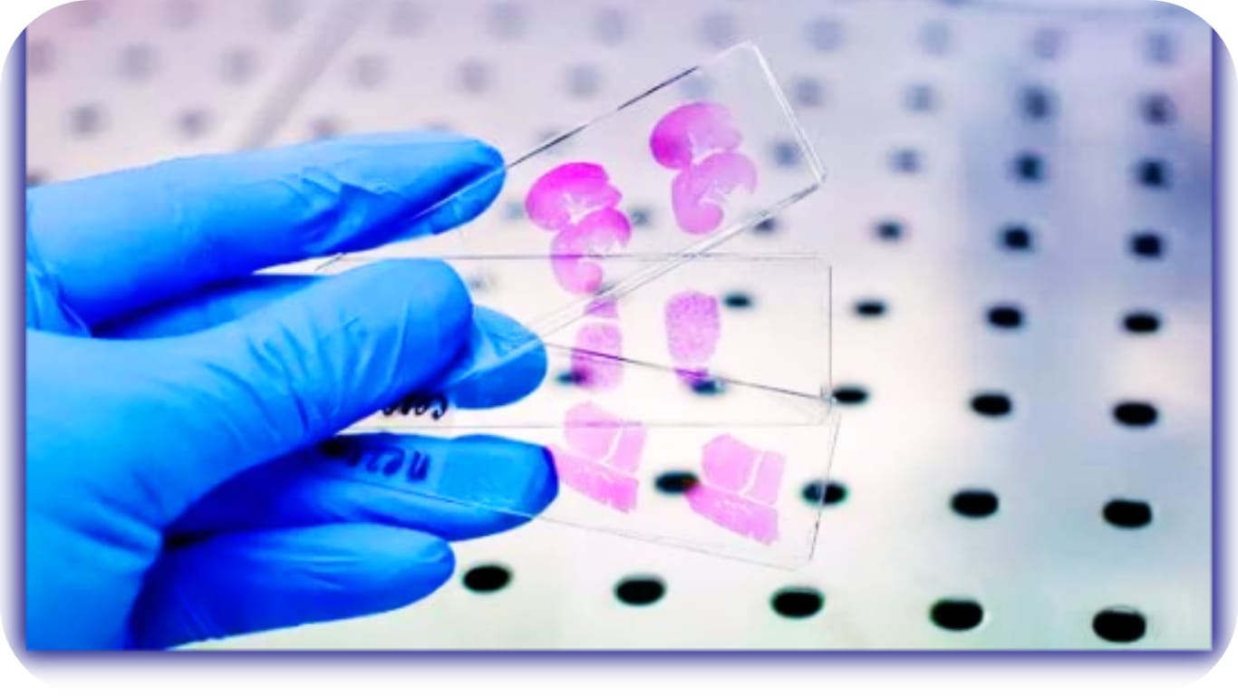
Immunohistochemistry (IHC) in Cancer
Tumor markers are molecules whose levels are considered as signals, symbols, or representatives of tumor cells, and increased in cancerous conditions. Normal cells express most of the tumor markers like tumor cells. By present, most tumor markers are found to be proteins, and some patterns of gene expression and DNA changes are also considered to be tumor markers. More than 20 tumor markers have been developed for clinical use. One kind of cancer is normally featured with one or more tumor markers. According to immunohistochemistry (IHC) visualized tumor markers such as enzymes, oncogenes, tumor-specific antigens, tumor suppressor genes and tumor proliferation markers, doctors can efficiently predict oncogenesis and diagnose a cancer as benign or malignant, determine the stage and the grade of a cancer.
Immunohistochemistry in Lung Cancer
Immunohistochemistry represents an important complementary tool for the routine diagnosis of lung cancer and for the identification of the different histological types and prognostic factors. Its purpose is to categorize patients in order to ensure appropriate and specific treatment, as well as to identify tumors at higher risk of recurrence and fatal outcomes. The essential immunohistochemistry panel recommended for the diagnosis and prognosis includes expression of the following markers: CK7; CK20; COX-2; TTF-1, chromogranin; synaptophysin, CD56; PSA; CA125; p53; c-erbB-2; MMP-9; and VEGF.
Reference: Role of immunohistochemistry in the diagnosis of lung cancer, Vera Luiza Capelozzi.
Immunohistochemistry is playing an increasing role in the modern pathology of breast disease. Some of the diagnostic uses of IHC include differentiating UDH from ADH/LG-DCIS, ruling out microinvasion, distinguishing invasive carcinoma from pseudoinvasive lesions, identifyingbreast cancer histologic subtype and molecular phenotype, and confirming the breast as the primary site in metastatic carcinoma. In addition, immunohistochemical markers are useful for estimating prognosis and predicting therapy response. The best approach to the use of immunohistochemical markers is to couple them with standard hematoxylin-eosin histology and to use panels of markers.
Reference: Application of Immunohistochemistry to Breast Lesions, I-Tien Yeh, MD; Carolyn Mies, MD
Immunohistochemistry in Prostate Cancer
The use of IHC as an auxiliary in the diagnosis of adenocarcinoma is a common practice in uropathology, and the use of antibodies against p63 and high molecular weight cytokeratin has been recommended as adjuncts in confirming prostatic carcinoma in doubtful cases. Although basal cell markers, such as 34βE12 and p63 antibodies are useful for identifying basal cells, several benign mimickers of PC, such as atrophy, atypical adenomatous hyperplasia, nephrogenic adenoma, and mesonephric hyperplasia, can stain negatively with these markers. In addition, with patients being submitted to prostate biopsy with lower PSA levels and with larger numbers of cores being taken in each biopsy session, concern that patients are being overtreated is common.
Reference: The Use of Immunohistochemistry for Diagnosis of Prostate Cancer, Katia R. M. Leite, et al
| SI | Department | Exam Name | MRP in BDT | Delivery Hour Normal |
| 1 | IMMUNOHISTOCHEMISTRY | ALK (IHC) | 8,000 | 168 |
| 2 | IMMUNOHISTOCHEMISTRY | CD - 117 [ IHC ] | 6,500 | 168 |
| 3 | IMMUNOHISTOCHEMISTRY | ER (Estrogen Receptor) | 5,000 | 168 |
| 4 | IMMUNOHISTOCHEMISTRY | IMMUNOHISTOCHEMISTRY EXAMINATION - HER2 | 5,000 | 168 |
| 5 | IMMUNOHISTOCHEMISTRY | IMMUNOHISTOCHEMISTRY FOR LYMPHOMA PANEL | 18,000 | 360 |
| 6 | IMMUNOHISTOCHEMISTRY | Immunohistochemistry Examination - ER+PR+HER2 | 12,000 | 168 |
| 7 | IMMUNOHISTOCHEMISTRY | Immunohistochemistry Examination - ER+PR+HER2+KI67 | 16,000 | 168 |
| 8 | IMMUNOHISTOCHEMISTRY | Immunohistochemistry Examination - Ki67 | 5,000 | 168 |
| 9 | IMMUNOHISTOCHEMISTRY | Immunohistochemistry Examination - Final Diagnosis | 18,000 | 168 |
| 10 | IMMUNOHISTOCHEMISTRY | PD - L1 | 16,000 | 192 |
| 11 | IMMUNOHISTOCHEMISTRY | PR (Progesterone Receptor) | 5,000 | 168 |
Cancer markers:
ALK gene rearrangements
Cancer types:
Non-small cell lung cancer and anaplastic large cell lymphoma
Tissue analyzed: Tumor
How used: To help determine treatment and prognosis
Alpha-fetoprotein (AFP)
Cancer types:
Liver cancer and germ cell tumors
Tissue analyzed: Blood
How used: To help diagnose liver cancer and follow response to treatment; to assess stage, prognosis, and response to treatment of germ cell tumors
Beta-2-microglobulin (B2M)
Cancer types:
Multiple myeloma, chronic lymphocytic leukemia, and some lymphomas
Tissue analyzed: Blood, urine, or cerebrospinal fluid
How used: To determine prognosis and follow response to treatment
Beta-human chorionic gonadotropin (Beta-hCG)
Cancer types:
Choriocarcinoma and testicular cancer
Tissue analyzed: Urine or blood
How used: To assess stage, prognosis, and response to treatment
BCR-ABL fusion gene
Cancer type:
Chronic myeloid leukemia
Tissue analyzed: Blood and/or bone marrow
How used: To confirm diagnosis and monitor disease status
BRAF mutation V600E
Cancer types:
Cutaneous melanoma and colorectal cancer
Tissue analyzed: Tumor
How used: To predict response to targeted therapies
CA15-3/CA27.29
Cancer type:
Breast cancer
Tissue analyzed: Blood
How used: To assess whether treatment is working or disease has recurred
CA19-9
Cancer types:
Pancreatic cancer, gallbladder cancer, bile duct cancer, and gastric cancer
Tissue analyzed: Blood
How used: To assess whether treatment is working
CA-125
Cancer type:
Ovarian cancer
Tissue analyzed: Blood
How used: To help in diagnosis, assessment of response to treatment, and evaluation of recurrence
Calcitonin
Cancer type:
Medullary thyroid cancer
Tissue analyzed: Blood
How used: To aid in diagnosis, check whether treatment is working, and assess recurrence
Carcinoembryonic antigen (CEA)
Cancer types:
Colorectal cancer and breast cancer
Tissue analyzed: Blood
How used: To check whether colorectal cancer has spread; to look for breast cancer recurrence and assess response to treatment
CD20
Cancer type:
Non-Hodgkin lymphoma
Tissue analyzed: Blood
How used: To determine whether treatment with a targeted therapy is appropriate
Chromogranin A (CgA)
Cancer type:
Neuroendocrine tumors
Tissue analyzed: Blood
How used: To help in diagnosis, assessment of treatment response, and evaluation of recurrence
Chromosomes 3, 7, 17, and 9p21
Cancer type:
Bladder cancer
Tissue analyzed: Urine
How used: To help in monitoring for tumor recurrence
Cytokeratin fragments 21-1
Cancer type:
Lung cancer
Tissue analyzed: Blood
How used: To help in monitoring for recurrence
EGFR mutation analysis
Cancer type:
Non-small cell lung cancer
Tissue analyzed: Tumor
How used: To help determine treatment and prognosis
Estrogen receptor (ER)/progesterone receptor (PR)
Cancer type:
Breast cancer
Tissue analyzed: Tumor
How used: To determine whether treatment with hormonal therapy (such as tamoxifen) is appropriate
Fibrin/fibrinogen
Cancer type:
Bladder cancer
Tissue analyzed: Urine
How used: To monitor progression and response to treatment
HE4
Cancer type:
Ovarian cancer
Tissue analyzed: Blood
How used: To assess disease progression and monitor for recurrence
HER2/neu
Cancer types:
Breast cancer, gastric cancer, and esophageal cancer
Tissue analyzed: Tumor
How used: To determine whether treatment with trastuzumab is appropriate
Immunoglobulins
Cancer types:
Multiple myeloma and Waldenström macroglobulinemia
Tissue analyzed: Blood and urine
How used: To help diagnose disease, assess response to treatment, and look for recurrence
KIT
Cancer types:
Gastrointestinal stromal tumor and mucosal melanoma
Tissue analyzed: Tumor
How used: To help in diagnosing and determining treatment
KRAS mutation analysis
Cancer types:
Colorectal cancer and non-small cell lung cancer
Tissue analyzed: Tumor
How used: To determine whether treatment with a particular type of targeted therapy is appropriate
Lactate dehydrogenase
Cancer type:
Germ cell tumors
Tissue analyzed: Blood
How used: To assess stage, prognosis, and response to treatment
Nuclear matrix protein 22
Cancer type:
Bladder cancer
Tissue analyzed: Urine
How used: To monitor response to treatment
Prostate-specific antigen (PSA)
Cancer type:
Prostate cancer
Tissue analyzed: Blood
How used: To help in diagnosis, assess response to treatment, and look for recurrence
Thyroglobulin
Cancer type:
Thyroid cancer
Tissue analyzed: Tumor
How used: To evaluate response to treatment and look for recurrence
Urokinase plasminogen activator (uPA) and plasminogen activator inhibitor (PAI-1)
Cancer type:
Breast cancer
Tissue analyzed: Tumor
How used: To determine aggressiveness of cancer and guide treatment
5-Protein signature (Ova1)
Cancer type:
Ovarian cancer
Tissue analyzed: Blood
How used: To pre-operatively assess pelvic mass for suspected ovarian cancer
21-Gene signature (Oncotype DX)
Cancer type:
Breast cancer
Tissue analyzed: Tumor
How used: To evaluate risk of recurrence
70-Gene signature (Mammaprint)
Cancer type:
Breast cancer
Tissue analyzed: Tumor
How used: To evaluate risk of recurrence